Nervous System

One critical element to maintaining homoeostasis is the ability of an organism to communicate the changes in the internal and external environments and to control the effects of any perturbations. The two systems of the body that share this responsibility are the nervous system and the endocrine system, each greatly influencing the effects of the other.
The nervous system differs from the endocrine system primarily with respect to the speed and duration of effects. The nervous system produces very rapid and transient effects, suited to respond to the rapid changes in the environment, while the endocrine system typically produces effects that develop more slowly and tend to have a prolonged effect, suited to long term environmental alterations. The nervous system has three basic functions: sensory, integrative, and motor.
The nervous system can be broken into two primary branches: the central nervous system (CNS), and peripheral nervous system (PNS). The CNS is comprised of the brain and spinal cord while the PNS consists of cranial and spinal nerves, and the resulting branches.
I. Neurons

Neurons conducting impulses from the PNS to the CNS are called sensory (afferent) while the neurons conducting impulses from the CNS to the PNS are called motor (efferent). The PNS is divided into a somatic and autonomic division, with somatic consisting of neurons conducting impulses to and from cutaneous and special receptors, and the skeletal muscular system. The autonomic nervous system (ANS) consists of neurons conducting impulses to visceral organs, and may be further divided into the sympathetic and parasympathetic divisions.
Most of the synthesis in the physiological processes of the neuron occur in the soma, which is often at some distance from the axon terminals. In order to move the synthesized chemicals (such as neurotransmitters or their precursors) to the site of their use, particles must be moved down the axons. Recycling of such chemicals after their use (recovered from the synaptic cleft by active transport) also requires their shipment up the axon to the soma for reuse. Both a slow and fast axoplasmic transport system exists, slow due to normal sarcoplasmic flow, and the fast system utilizing protein carriers and a microtubule system to move specific particles.
Neurons are classified by structure as:

- multipolar, with many dendrites extending from the soma and one axon.
- bipolar, with one main dendrite extending from the soma and one axon.
- unipolar, with one main process extending from the soma so that the soma appears to be "set off to the side." Both branches of the process have the characteristics of an axon. Neurons are also classified by function as sensory neurons if they carry impulses from the PNS to the CNS, motor neurons if they carry impulses to the CNS to the PNS, or interneurons (association) if they are not specifically motor or sensory in function.

An electrically excitable membrane is a membrane, which will conduct electric events. Membranes must carry a resting electric charge (resting membrane potential) in order for this phenomenon to occur. The existence of a selectively permeable membrane, specifically in it’s permeability to ions, will provide the electrical nature necessary for such events.
In neurons resting membrane potential is about 70mV with the intracellular fluid approximately 70mV more negative than the extra cellular fluid when the membrane is at rest. This electrical potential exists due to the differences in the concentration of the cations and anions on either side of the membrane. Extracellular fluid is rich in sodium and chloride ions, while the intracellular fluid is rich in potassium ions and organic anions of amino acids (in proteins) and phosphates. In particular, the distribution of sodium becomes a critical ion in determining the potential. The plasma membrane of neurons contains many potassium/sodium pumps which actively move sodium outside of the cell while actively moving potassium inside. At rest, the membrane allows a certain permeability to potassium, but it is practically impermeable to sodium. The total distribution of ions results in an imbalance of charges that can be measured as resting membrane potential.
Ion channels allow the diffusion of ions across the membrane and may either be "leaky" or "gated." Leaky channels are always open, but gated channels open and close in response to stimuli. Such channels are typically specific in the modality of the stimulus to which they respond. Four types of gated channels are known: voltage, chemical, mechanical, and light. Voltage channels open in response to changes in membrane potentials, chemical channels open in response to the binding of specific chemicals to receptors at the channel, mechanical channels open in response to vibration or pressure changes, light channels open in response to particular wavelengths of light. The ability of a membrane to open/close channels, allows for the propagation of electrical impulses, as dramatic changes in the membrane potential will occur when ions are allowed to diffuse across the membrane.
A gated potential is an alteration in the resting membrane potential due to the influx or efflux of ions across the membrane. This flow of ions occurs as ion channels are open, allowing the ions to follow their electrical or chemical gradients. These are localized events due to the normal loss of energy produced by resistance to electrical flow. A membrane potential change that produces less negativity across the membrane is called a depolarization and is typically produced by opening a sodium ion channel, which allows for the flow of sodium ions into the cell. A membrane potential change that produces more negativity across the membrane is called a hyperpolarization and is typically produced by opening chloride ion channels, which allows the flow of chloride ions into the cell or by opening additional potassium channels which allow the flow of potassium ions out of the cell. Typically, graded potentials occur primarily on the soma and dendrites of neurons.
Neuron Behavior
In order to produce electrical nerve impulses, neurons must be triggered by a stimulus, which is anything inside or outside the body that evokes a physical or psychological response. A neurons capacity to respond to stimulus is known as excitability. Electrical nerve impulses may also be blocked or inhibited by some neurotransmitters or by drugs at the synapse. To fire a neuron, a stimulus must convert the electrical charge on the inside of the cell membrane from negative to positive. The nerve impulse travels down the axon to a synaptic knob and triggers the release of chemicals that may stimulate a response in target cells. Neurofilaments act as scaffolding to help give the nerve cell it’s shape. Cell membranes convey electrical impulses away from the cell body. Microtubules are thought to help in the transport of neurotransmitter molecules to the synaptic membrane. Synaptic vesicles are sacs that contain molecules of neurotransmitters that are drawn towards synaptic cleft by calcium ions. The neurotransmitter combines with protein receptors sites on the target cell membrane, which then becomes permeable to specific ions. Each knob at the end of an axon terminal fiber lies close to the neuron cell body, its axons, or dendrites, another synaptic knob, or a muscle fiber. Neurotransmitter molecules are released from the vesicles into the synaptic cleft where they influence impulse transmission. Excitation occurs when enough positive sodium ions have passed through channels in the membrane to change the charge on the inside of the cell membrane from negative to positive.
The level at which a stimulus begins to transmit an electrical impulse is called a threshold. If the stimulus is too weak or below the threshold, there is only a brief local response in the membrane. If, however, the threshold is reached, the impulse travels along the entire length of the fiber. The speed of transmission can vary: fibers that are cold (as when ice is applied in order to dull pain, those with small diameters, those without myelin sheaths conduct impulses more slowly.)
In a neurons resting state, when no impulse is being transmitted, Na+ ions diffuse from the inside of the cell membranes at a continuous rate. The inner membrane of the cell is thus negatively charged.
When stimulated by a nerve impulse, positively charged ions in the fluid outside of the membrane cross the cell membranes. At these local sites, the electrical charge on the inside of the cell membrane changes from negative to positive.
This localized reversal of charge across the membrane stimulates similar changes in the subsequent membrane segments. The electrical impulse continues down the axon as it passes along previous segments of the membrane reverting back to the "inside-negative" state.
The impulse reaches the synaptic cleft. When a neurotransmitter is released from vesicles in the synaptic knob, it crosses the synaptic cleft and stimulates the muscle fibers to contract.
Peripheral nerve fibers that are crushed or partially cut may slowly regenerated if the cell body and the segments of myelin sheath remain continuous. Regeneration does not occur in nerves in the brain or spinal cord, instead injured nerve fibers are wrapped in scar tissue and inactivated.
When a nerve fiber just beyond an injury and farthest from the cell body no longer receive vital proteins and enzymes, it begins to degenerate and the myelin sheath becomes hollow. The undamaged neurons cell body stimulates the growth of several nerves sprouts in the remaining portion of the fiber. One of these sprouts may eventually find it’s way through the empty but intact myelin sheath.
Growing at a rate of about 1.5mm per day, the new nerve fiber reaches its previous connection. Function and sensation are slowly restored and unused nerve sprouts degenerate. During inhibition or blockage of electrical impulse channels that are sensitive to chloride or potassium ions may open rather than channels that are sensitive to sodium. Positive potassium ions escape from the target cell or else negative chloride ions permeate the cell membrane. In both cases, the electrical charge inside of the target cell membrane stays negative and the neurons can’t be fired and the nerve impulse is inhibited.
II. Central Nervous System
Located in the skull, the brain is composed of more than 12 billion neurons and 50 billion supporting glial cells but it weighs less than 3 pounds. With the spinal cord, the brain monitors and regulates many unconscious bodily processes such as heart rate and coordinates most voluntary movement. Most important is the site of consciousness and of all the intellectual functions that allow humans to think and create.
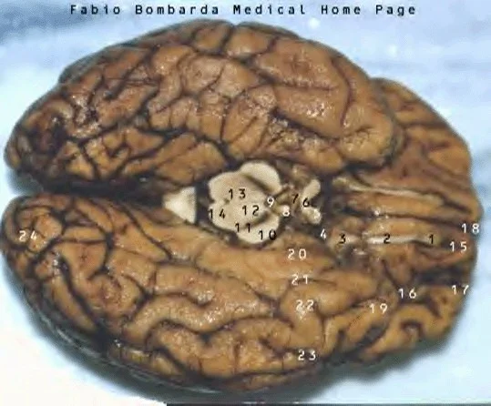
The most obvious feature about the cerebrum, the largest part of the brain, is its heavily folded surface, the pattern of which is different in each human being. The grooves are called sulci when shallow and fissures when deep. Fissures and some large sulci outline specific functional areas that are called lobes. A ridge on the brains surface is a called a gyrus. A longitudinal fissure divides the brain into two halves, called hemispheres, that communicate with each other. Speech production, the elaboration of thought and emotions, skilled movements are controlled by neurons found in the frontal lobe of the brain. The recognition of specific tones and loudness takes place in the temporal lobe. This area also plays a role in memory storage. Higher intellectual function such as memory and the interpretation of sensory impulses are assisted by the complex network of neurons located in the cerebral cortex. Neurons of the cerebellum link with other regions of the brain and the spinal cord, facilitating smooth, precise movement and controlling balance and position. It also plays a role in speech. The occipital lobe detects and interprets visual images. A variety of bodily sensations such as touch, pressure, pain, and temperature are both perceived and interpreted in the parietal lobes. The entire cerebrum is covered by a layer of gray matter about 2 to 6 mm thick. Underneath is the brains white matter as well as islands of gray matter.
Within the center of the brain lies the thalamus which is the brains information relay station. Surrounding the thalamus is a group of structures, the limbic system that is involved in survival behavior and emotions, such as rage and fright. Closely linked with the limbic system is the hypothalamus, which has overall control of the bodies automatic processes.
The brain's gray matter is made up of groups of neuron cell bodies. White matter, by contrast, is composed mainly of the myelin covered axons of nerve fibers that extend from the neuron cell bodies. Fatty myelin sheaths insulate the axons and increase the transmission speed of the nerve impulses. Basogangliar masses or nuclei of gray matter found deep in the brain that help control movement, sequences such as walking. The corpus callosum is the largest of several bundles of nerve fibers called commissures that connects specific areas of the two hemispheres in the brain.
Myelinated fibers organized into so called projection tracts transmit impulses to and from the cerebral cortex to the lower brain and spinal cord. These nerve tracts pass through a communicational link called the internal capsule, a compact band of fibers that intersect the corpus collosum. After passing through the upper part of the brain stem, projection fibers fan out and extend to the cerebral cortex forming the cornii radii.
The thalamus is a relay station that sorts, interrupts, and directs sensory signals received from both the spinal cord and midbrain to the cerebral cortex and to appropriate sites in the cerebrum. The brain stem contains centers that regulate several functions that are vital to survival: these include blood pressure, heart beat, respiration, digestion, and certain reflex reactions such as swallowing and vomiting.
The soft tissue of the brain floats within the bony casing of the skull in a watery medium, which also flows around the spinal cord, called cerebral spinal fluid (CSF). This colorless liquid, which is renewed on a continuous basis, is produced inside the ventricles or chambers of the brain. The fluid contains glucose, necessary to provide energy for cell function of the brain and spinal cord, as wells as proteins and lymphocytes that guard against infection. Cerebral spinal fluid is produced in clusters of thin walled capillaries called chorideplexes that line the walls of the ventricles.
Cerebral spinal fluid moves from the lateral ventricles into the 3rd and 4th ventricles. It then flows up the back of the brain, down around the spinal cord, and up around the front of the brain. After circulating cerebral spinal fluid is reabsorbed into blood through arachnoid granulations, projections from arachnoid layer of the meninges that connect via the veins, via the venous sinus. Each of the two lateral ventricles, one in each hemisphere has a front and longer back horn, which extend from the center of the brain. These ventricles have an appearance of a large X.
The solid bones of the skull may fracture if struck sufficiently hard. However, the CFS within the skull absorbs and disperses excessive mechanical forces that might otherwise cause serious injury to the brain. Analysis of its chemicals constitutes and the pressure of its flow offers vital clues to the diagnoses of many disorders of the brain, such as meningitis. Fluid produced in the lateral ventricles drains via the interventricle form and to the 3rd ventricle close to the thalamus. It then flows through the cerebral aqueduct and into the 4th ventricle, which is located in front of the cerebellum. Circulation is aided by the pulsations of the cerebral arteries. Aided by ventricular movement, the fluid flows downward along the back of the spinal cord, in the central canal and upper along the ____ of the cord.
Three membranes known as the meninges cover the brain. Lining the inside of the skull is the outer most membrane, the dura mater, which contains veins and arteries that nourish cranial bones. The middle layer is known as the arachnoid (spider-like) which consists of a type of web like elastic connective tissue. Next to the surface of the cerebral cortex is the pia mater. Between this delicate innermost layer of the arachnoid, is the subarachnoid space, which contains cerebral spinal fluid as well as blood vessels.
Although the brain accounts for only about 2% of the total weight of body, it requires 20% of the bodies blood, oxygen and glucose are transported by blood, without these essential elements, brain function quickly deteriorates and dizziness and confusion and loss of consciousness may occur. Within only 4 to 8 minutes of oxygen deprivation, brain damage or death results.
Two front and two back arteries join up at the base of the skull to form an arterial wing called the circle of Willis. From this point, branching blood vessels provide the brain with oxygenated blood. A controlled molecular flow to the brain is essential for stable brain function. Endothelial cells that are located in the capillary walls create an almost impassable layer. In addition, capillaries are also wrapped within the fibers of the protective neurons (astrocytes). Oxygen, water, glucose, and relatively small molecules pass through this two layer barrier easily, but many drugs and chemicals cannot pass through at all.
Brain growths most part of the embryonic life occurs much more rapidly than the development and growth of the limbs or internal organs. From small clusters of tissue, highly specialized areas of brain function emerge. At 3 weeks, a tube of neuro tissue develops along the back of the embryo. These bulges called the primary vesicles develop into the main divisions of the brain. At 7 weeks, the neuro tube flexes and cranial nerves sprout from the hindbrain. Bulges form on the forebrain, one of which will develop into the cerebrum. At 11 weeks the hindbrain separates into the cerebellum, the pons, and the medulla. The forebrain develops further into the cerebrum, and starts to grow back over the hindbrain. At birth, as the cerebrum enlarges to become the largest part of the body, folding of the cerebral cortex occurs. Every individual has a unique folding pattern.
Spinal Cord
The spinal is a cable about 17 inches in length which extends from the brain stem to the lumbar part of the back. A slightly flattened cylinder is about as wide as a finger for most of its length, tapering into a thread like tail. Through 31 pairs of spinal nerves, the spinal cord is connected to the rest of the body and relays information received via these nerves about it’s internal and external environment to and from the brain.
The spinal cord consists of two types of tissue. The inner core is gray matter made up of neuron cells bodies, unmyelinated axons, glial cells, and blood vessels. It contains cell bodies of motor neurons that bring about voluntary reflex movements and control internal functions. Outer white matter is composed of tracts of myelinated axons that relay impulses to and from the spinal cord and specific areas of the brain. Cerebral spinal fluid fills the central canal and provides nourishment to nerve cells. Three layers of connective tissue called meninges protect the spinal cord. Additional protection is provided by cerebral spinal fluid circulating in the subarachnoid space. Impulses about bodily sensations are carried by sensory nerve fibers. These converses the form sensory reaches at the back of the spinal cord. Impulses are then conveyed in the cord to the brain via fiber tracts. Each spinal nerve has a dorsal or posterior sensory root ganglion, which is a cluster of nerve cell bodies. Bundles of fibers called motor nerve roots emerge from the front of the spinal cord. Motor nerve fibers conduct impulses to the voluntary skeletal muscles. Separate nerves controlling the involuntary processes form the autonomic nervous system. The spinal nerves run through gapes between adjacent vertebrate and enter the back and front of spinal cord as spinal nerve roots.